




Ultrasound In Pregnancy
Home » Ultrasound in Pregnancy: Safe, Essential Scans Explained | The Ova Fertility and Women Care
Why The Ova Care Is Trusted as the Best Fertility and Maternity Hospital

Personalized Support

High Success Rate

24/7 Availablity

5000+ Successful Pregnancies

16+ Years of Experienced Experts

Affordable & Ethical Treatment
Ultrasound In Pregnancy
Ultrasound in pregnancy is one of the most important tools used to monitor the baby’s development and the mother’s health during antenatal care. It uses high-frequency sound waves to create images of the baby, uterus, placenta, cervix, amniotic fluid, and surrounding structures. Unlike X-rays, pregnancy ultrasound does not use radiation. When performed for medical reasons by trained professionals, it is considered safe and valuable throughout pregnancy.
At The Ova Fertility & Women Care, ultrasound in pregnancy in Thane is used to confirm early pregnancy, check fetal heartbeat, calculate gestational age, assess fetal growth, evaluate the placenta, measure amniotic fluid, identify selected structural concerns, monitor fetal wellbeing, and help plan care as pregnancy progresses. Each scan has a purpose. Some scans are routine, while others are advised only when the doctor needs additional information.
A pregnancy scan should never be treated as only a picture of the baby. It is a clinical assessment that must be interpreted in context. The report is most useful when it is reviewed alongside maternal symptoms, blood tests, previous scans, fetal movements, and antenatal history.
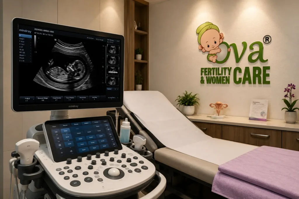
What Is Pregnancy Ultrasound?
Pregnancy ultrasound, also called prenatal ultrasound, obstetric ultrasound, fetal ultrasound, pregnancy scan, or antenatal ultrasound, is an imaging test used during pregnancy to assess the baby and pregnancy-related structures. The body is probed with sound waves using a handheld transducer. These waves reflect back from tissues and are converted into images on a screen.
Most pregnancy scans are performed through the abdomen. In early pregnancy, a transvaginal scan may sometimes be advised because the pregnancy is very small and clearer images may be needed. This is especially helpful when confirming early viability, checking the pregnancy location, or evaluating bleeding or pain in early pregnancy.
Ultrasound during pregnancy can provide information that physical examination alone cannot. It helps the doctor see whether the pregnancy is inside the uterus, whether the baby’s heartbeat is present, whether growth is appropriate, where the placenta is located, how much amniotic fluid surrounds the baby, and whether further assessment is needed.
Is Ultrasound Safe During Pregnancy?
This is one of the most common questions mothers ask. Ultrasound uses sound waves, not radiation. When used appropriately for medical purposes, it has not been shown to harm the developing baby. It is used worldwide as a standard part of pregnancy monitoring.
However, ultrasound should be performed with medical purpose and proper technique. The scan frequency should be decided by the doctor based on pregnancy stage, previous reports, maternal health, fetal growth, and symptoms. Extra scans are useful when they answer a clinical question, but unnecessary scanning without indication should be avoided.
At Ova Care, ultrasound is used responsibly. The mother is told why the scan is being done, what it is expected to assess, and when follow-up may be needed.
Why Ultrasound Is Done During Pregnancy
Ultrasound in pregnancy can help confirm pregnancy, check location, identify heartbeat, estimate gestational age, calculate the expected delivery date, assess fetal development, check the placenta, measure amniotic fluid, evaluate fetal position, monitor growth, and guide delivery planning when required.
In early pregnancy, ultrasound can help identify an ectopic pregnancy, where the pregnancy is located outside the uterus. This can be dangerous if not diagnosed early. Ultrasound can also help evaluate bleeding, pain, uncertain dates, or miscarriage concerns.
In later pregnancy, fetal growth ultrasound and fetal wellbeing assessment help the doctor understand whether the baby is growing as expected. Ultrasound can also assess placental position, fluid levels, baby’s position, and selected signs that may require closer monitoring.
First Trimester Ultrasound
The first trimester ultrasound is usually performed in early pregnancy. It may be advised around 6 to 8 weeks to confirm the location of the pregnancy, detect the heartbeat, check whether there is one baby or more than one, and estimate gestational age. This scan is especially useful if menstrual dates are uncertain, periods are irregular, or pregnancy followed fertility treatment.
An early pregnancy scan can also help evaluate bleeding, cramping, pain, or suspected ectopic pregnancy. If the scan is done too early, the heartbeat may not yet be visible. In such cases, the doctor may advise repeat ultrasound after a suitable interval rather than giving a premature conclusion.
A pregnancy dating scan helps establish the expected delivery date more accurately, especially in early pregnancy. Accurate dating is important because later decisions about growth, screening, and delivery timing depend on the correct gestational age.
NT Scan and First Trimester Screening
Between 11 and 14 weeks, a scan may be advised to assess nuchal translucency, commonly called NT. This is a measurement of fluid thickness behind the baby’s neck. It is usually interpreted along with blood tests and maternal age to assess the risk of certain chromosomal conditions.
The NT scan can also provide early information about fetal development, heartbeat, gestational age, and sometimes early structural assessment.This is a screening test, not a diagnosis. If the result suggests increased risk, the doctor may discuss further testing options.
At Ova Care, first trimester screening is explained clearly so parents understand what the test can and cannot tell them. Anxiety often comes from unclear reports; a calm explanation helps families make informed decisions.
Second Trimester Ultrasound
The second trimester ultrasound is one of the most detailed scans in pregnancy. The anomaly scan, usually performed around 18 to 22 weeks, evaluates fetal anatomy. It checks major structures such as the brain, spine, heart, abdomen, kidneys, limbs, face, stomach, bladder, and other visible organs. It also assesses placenta location, amniotic fluid, fetal growth parameters, and cervical length when indicated.
The anomaly scan cannot detect every possible condition, but it is an important fetal development assessment. Some findings may require repeat imaging, fetal echo, specialist opinion, or follow-up scans. Sometimes the baby’s position makes it difficult to see all structures clearly, and the mother may be asked to return for another scan.
This scan is medically important and emotionally significant. Many parents feel excited, but they may also feel nervous. The role of the care team is to perform the scan carefully, explain findings responsibly, and guide the next step if anything needs review.
Third Trimester Ultrasound
Third trimester ultrasound is used to assess fetal growth, amniotic fluid, placenta, fetal position, and overall wellbeing. It may be advised around the later weeks of pregnancy, depending on clinical need. Some mothers need only routine growth assessment, while others may need more frequent scans if the doctor wants closer observation.
A fetal growth scan estimates baby weight using measurements such as head size, abdominal circumference, and thigh bone length. It also helps compare growth with expected values for gestational age. Growth assessment is important because both poor growth and excessive growth may influence monitoring and delivery planning.
The third trimester scan may also show whether the baby is head-down, breech, or in another position. This information is useful closer to delivery. Placental location is reviewed again if earlier scans showed a low-lying placenta.
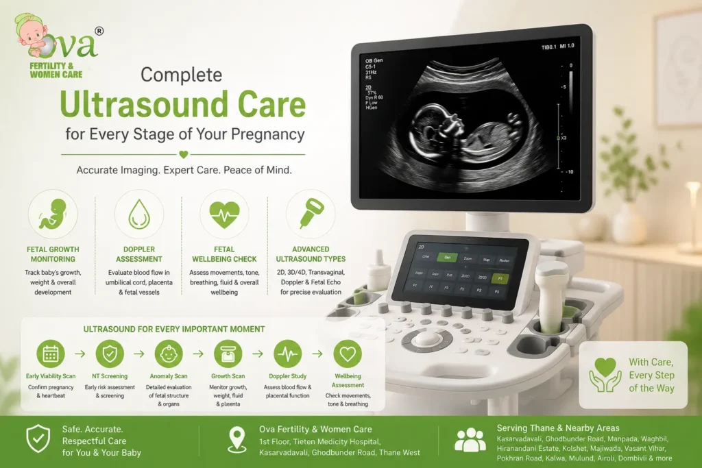
Fetal Growth Ultrasound
Fetal growth ultrasound is used to assess whether the baby is growing appropriately. It helps track growth patterns over time rather than relying on one measurement alone. If the baby appears smaller or larger than expected, the doctor may advise repeat scans, Doppler assessment, blood tests, or closer antenatal review.
Growth scans usually include fetal measurements, estimated fetal weight, amniotic fluid assessment, placenta review, and fetal movement observation during the scan. These details help the doctor decide whether the pregnancy can continue with routine monitoring or whether closer follow-up is needed.
The use of growth ultrasounds is particularly useful when clinical examination suggests a size difference, maternal weight limits abdominal assessment, prior pregnancy history raises concerns, fetal movements change, and medical conditions require monitoring.
Doppler Ultrasound During Pregnancy
Doppler ultrasound evaluates blood flow in selected vessels. In pregnancy, it may be used to assess blood flow through the umbilical cord, placenta-related circulation, or fetal vessels when clinically indicated. Dopplers are not required for every routine pregnancy scan, but they can provide valuable information on fetal growth, placenta function, and wellbeing.
A Doppler report helps the doctor understand whether the baby appears to be receiving adequate blood supply. If the findings are abnormal, the doctor may advise closer monitoring, repeat scans, additional tests, or delivery planning depending on gestational age and overall condition.
Doppler findings should always be interpreted by the treating doctor. One number alone should not be read in isolation.
Fetal Wellbeing Assessment
Fetal wellbeing assessment may include ultrasound evaluation of fetal movements, tone, breathing movements, amniotic fluid, and sometimes fetal heart monitoring when needed. These assessments are used when the doctor wants to check how the baby is doing inside the uterus.
A biophysical profile may be advised in selected cases. It combines ultrasound observations to give information about fetal wellbeing. Your current Ova page already mentions biophysical profile and computerized fetal heart monitoring as tools used closer to delivery in selected situations. These assessments help guide pregnancy decisions when reassurance or closer observation is needed.
It is important for mothers to stay aware of their fetal wellbeing. Reduced fetal movements should always be reported promptly.
Types of Pregnancy Ultrasound
Abdominal ultrasound is the most common type used during pregnancy. Gel is applied to the abdomen, and the probe is moved over the skin to obtain images. It is painless, though pressure may feel uncomfortable if the bladder is full or the abdomen is tender.
Transvaginal ultrasound may be used in early pregnancy or when clearer pelvic images are needed. A slim probe is placed inside the vagina with appropriate hygiene and privacy. It is often more accurate in early weeks because the pregnancy is very small.
Doppler ultrasound assesses blood flow. Fetal echo is a specialized scan of the baby’s heart and may be advised if there are risk factors or scan findings that need detailed cardiac assessment. 2D ultrasound is the standard medical imaging format. 3D or 4D scans may show surface images, but they should not replace medically indicated 2D assessment.
What Each Pregnancy Scan Checks
An early pregnancy scan checks pregnancy location, heartbeat, number of embryos, gestational age, and early concerns such as ectopic pregnancy or bleeding-related findings. A dating scan helps confirm the expected date of delivery. First trimester screening assesses early fetal markers and risk indicators.
The anomaly scan checks fetal structure and organ development. A fetal growth scan assesses size, estimated weight, amniotic fluid, placenta, and growth pattern. Doppler checks selected blood-flow patterns. Late pregnancy scans may assess baby position, fluid, placenta, and growth before delivery planning.
Each scan has a different purpose. Repeating the same scan without reason does not necessarily add value. The doctor decides timing based on pregnancy stage and clinical need.
How to Prepare for a Pregnancy Scan
Preparation depends on the type of scan and pregnancy stage. For early abdominal scans, the doctor may ask for a comfortably full bladder because it can improve visibility. For transvaginal scans, the bladder is usually emptied. For later pregnancy scans, special preparation is often not required unless instructed.
Wear comfortable clothing that allows the abdomen to be exposed easily. Carry previous scan reports, blood reports, antenatal file, medication list, and doctor’s notes. If you have symptoms such as bleeding, pain, leaking fluid, fever, or reduced fetal movements, tell the team before the scan.
Most scans are not painful. The gel may feel cold, and pressure from the probe may be mildly uncomfortable. If anything hurts, inform the sonographer or doctor.
Understanding Your Report
A pregnancy ultrasound report may include gestational age, fetal heartbeat, fetal measurements, estimated fetal weight, placenta location, amniotic fluid, fetal position, cervix assessment when relevant, and comments about visible anatomy depending on the scan type.
The person performing the scan may not always explain every detail immediately. The treating doctor should review the report in the context of your pregnancy. If the baby’s position prevents clear views, a repeat scan may be advised. If a finding is uncertain, follow-up imaging may be needed.
A report should not create panic without explanation. Some findings are minor and need only observation. Others need additional assessment. At Ova Care, scan findings are discussed in practical language so parents understand the next step.
Limitations of Ultrasound
Ultrasound is valuable, but it cannot detect every condition. Some abnormalities may not be visible due to fetal position, maternal body habitus, gestational age, low fluid, movement, or technical limitations. Some conditions develop later and may not be present at an earlier scan. Some genetic, metabolic, neurological, or developmental conditions cannot be ruled out by ultrasound alone.
Anomaly scan reduces uncertainty, but it does not guarantee that the baby has no health issues. Similarly, a normal growth scan is reassuring for that visit, but continued antenatal monitoring remains important.
Responsible ultrasound counselling includes explaining both the benefit and the limitation of imaging.
When Extra Scans May Be Needed
Additional scans may be advised if the mother has bleeding, pain, reduced fetal movements, uncertain dates, previous pregnancy concerns, growth differences, low or high fluid, low-lying placenta, suspected fetal anomaly, multiple pregnancy, medical conditions, abnormal screening results, or post-date monitoring needs.
Extra scans are not meant to worry the mother. They help the doctor observe the baby more closely and decide whether care should continue routinely or change. The number of scans is individualized.
At Ova Care, the scan plan is based on medical need, not fear.
Emotional Side of Pregnancy Ultrasound
For many parents, seeing the baby on screen is a memorable moment. It can bring joy, relief, and a stronger emotional connection. It can also bring anxiety, especially while waiting for heartbeat confirmation or anomaly scan results.
The care team should recognize both emotions. A pregnancy scan is a medical examination, but it is also a sensitive family moment. Calm communication, privacy, respectful explanation, and clear follow-up make a significant difference.
Why Choose Ova Care for Ultrasound in Pregnancy?
The Ova Fertility & Women Care provides ultrasound in pregnancy in Thane with antenatal coordination, fetal development assessment, pregnancy imaging, growth monitoring, and doctor-led interpretation. Your current page already highlights the importance of early viability scans, NT screening, anomaly scan, growth monitoring, Doppler assessment, amniotic fluid evaluation, and late pregnancy scan planning. The rewritten page builds on those strengths with clearer structure and patient-focused explanation.
Fertility Treatments
Let's Meet Up
contact
Frequently Asked Questions
Yes. Pregnancy ultrasound uses sound waves and does not use radiation. When performed for medical reasons by trained professionals, it is considered safe during pregnancy.
The first ultrasound may be done around 6 to 8 weeks to confirm pregnancy location, heartbeat, number of embryos, and gestational age.
A dating scan estimates gestational age and expected delivery date. It is most accurate when performed in early pregnancy.
An anomaly scan is usually performed around 18 to 22 weeks to assess fetal anatomy, major organs, placenta, fluid, and growth markers.
A fetal growth scan measures baby size, estimated weight, amniotic fluid, placenta, and growth pattern. In addition, it gives you a sense of how your baby is growing.
A full bladder may be advised for early abdominal scans. For transvaginal scans, the bladder is usually emptied. Follow the instruction given before your appointment.
Yes. Transvaginal ultrasound is commonly used in early pregnancy when clearer images are needed. It should be performed gently and only when clinically appropriate.
No. Ultrasound can detect many structural concerns, but it cannot identify every condition. Some problems may develop later or may not be visible on scan.
Repeat ultrasound may be needed if the baby’s position limits views, dates are uncertain, growth needs monitoring, fluid or placenta needs reassessment, or the doctor wants follow-up.
Report timing depends on the scan type and center workflow. The treating doctor should review the report and explain what the findings mean for your pregnancy.
Related Searches:
Intrauterine Insemination (IUI) Treatment in Thane, Pre-Implantation Genetic Testing in Thane, Blastocyst Culture And Transfer Clinic in Thane, Laser Assisted Hatching Treatment in Thane, Egg/Sperm/Embryo Cryopreservation Freezing, Endoscopy In Fertility Treatments, Pregnancy Care, Birthing, Ultrasound In Pregnancy, High Risk Pregnancy Treatment Care With ICU Facility, Laparoscopic Surgery, Hysteroscopic Surgery, Menopause, Puberty Adolescence Treatment, Oncofertility,Male Infertility Treatment in Thane, best ivf hospital, ivf specialist, ivf doctor in thane, fertility treatment in thane, dr. snehal kohale ivf specialist
Our Latest Articles

Unexplained Infertility: What It Means When Routine Tests Are Normal
For many couples, receiving normal fertility test reports but still not achieving pregnancy can feel confusing and frustrating. When standard evaluations do not identify a clear reason for difficulty conceiving, doctors may describe the situation as unexplained infertility. This does not mean that there is no reason behind the fertility
Thyroid and Infertility: How TSH Can Affect Ovulation and Pregnancy Planning
When couples are planning a pregnancy, doctors often evaluate different hormones that play a role in reproductive health. Among these, thyroid hormones are important because they influence metabolism, menstrual cycles, ovulation, and pregnancy preparation. The connection between thyroid and infertility is mainly related to how thyroid hormone imbalance can affect

High Prolactin and Infertility: How Prolactin Can Affect Ovulation
When couples are trying to conceive, doctors often evaluate several hormones that influence reproductive health. One of these important hormones is prolactin. While prolactin is best known for its role in breast milk production, abnormal levels can also affect reproductive hormones and ovulation. The connection between high prolactin and infertility