




Hysteroscopic Surgery Treatment in Thane
Home » Hysteroscopic Surgery in Thane | Ova Fertility and Women Care
Why The Ova Care Is Trusted as the Best Fertility and Maternity Hospital

Personalized Support

High Success Rate

24/7 Availablity

5000+ Successful Pregnancies

16+ Years of Experienced Experts

Affordable & Ethical Treatment
Hysteroscopic Surgery Treatment in Thane
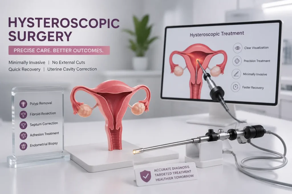
Hysteroscopic surgery is a minimally invasive uterine procedure used to examine and treat problems inside the uterus without any external abdominal cut. At Ova Fertility & Women Care, hysteroscopic surgery treatment in Thane is offered for women who need precise diagnosis or correction of intrauterine conditions such as endometrial polyps, submucous fibroids, uterine septum, intrauterine adhesions, abnormal bleeding, retained tissue, or suspected cavity abnormalities.
The uterus has an inner space called the uterine cavity. This cavity is lined by endometrium and plays an important role in menstruation, reproductive health, and overall gynecological wellbeing. Some conditions inside this cavity may not be fully understood through routine examination or even standard imaging. Hysteroscopy allows the gynecologist to directly view the inside of the uterus using a thin camera system called a hysteroscope. The image is seen on a monitor, allowing accurate assessment and targeted treatment.
Unlike laparoscopy, hysteroscopy does not involve cuts on the abdomen. The hysteroscope is passed through the vagina and cervix into the uterus. This no-cut approach makes hysteroscopic uterine surgery especially useful for selected women who need intrauterine correction with shorter recovery and less tissue trauma.
What Is Hysteroscopic Surgery?
Hysteroscopic surgery, also called operative hysteroscopy, therapeutic hysteroscopy, uterine endoscopy surgery, or intrauterine surgery, is a procedure in which a gynecologist uses a hysteroscope to diagnose or treat conditions inside the uterine cavity. A fluid, commonly saline, is used to gently expand the cavity so the doctor can see the uterine lining clearly.
A diagnostic hysteroscopy is performed mainly to inspect the inside of the uterus and confirm a finding. Operative hysteroscopy is performed when treatment is needed during the same sitting. Through the hysteroscope, fine instruments may be used to remove polyps, cut adhesions, resect a septum, remove selected fibroid tissue, take biopsy, or correct cavity abnormalities.
The main advantage is direct visualization. Instead of depending only on indirect images, the surgeon can see the abnormality and treat it precisely. This is why hysteroscopic treatment is considered valuable for uterine cavity correction.
When Is Hysteroscopic Surgery Advised?
Hysteroscopic surgery may be advised when symptoms or reports suggest a problem inside the uterus. Common reasons include heavy menstrual bleeding, irregular bleeding, bleeding between periods, prolonged periods, postmenopausal bleeding, suspected endometrial polyp, submucous fibroid, uterine septum, intrauterine adhesions, retained products, thickened endometrium, misplaced intrauterine device, or need for endometrial biopsy.
Some women are advised hysteroscopy after ultrasound, saline sonography, HSG, or MRI suggests a cavity abnormality. Others may need the procedure because symptoms continue despite apparently normal reports. Direct visualization can sometimes identify findings that are difficult to confirm on routine scans.
Hysteroscopic surgery should always be recommended with a clear purpose. The patient should know whether the goal is diagnosis, biopsy, polyp removal, fibroid resection, adhesion treatment, septum correction, or another specific uterine cavity procedure.
Diagnostic Versus Operative Hysteroscopy
Diagnostic hysteroscopy is used to inspect the uterine cavity. The doctor checks the endometrium, uterine shape, tubal openings, polyps, fibroids, adhesions, septum, inflammation, retained tissue, or abnormal areas that may need biopsy. It may be short and may not require extensive anesthesia in selected cases.
Operative hysteroscopy is treatment-focused. If a polyp, small submucous fibroid, septum, adhesion, or retained tissue is seen, the gynecologist may treat it using specialized instruments. Operative procedures may require anesthesia, more time, and closer post-procedure observation depending on complexity.
The distinction matters because recovery, fasting instructions, anesthesia, consent, and treatment planning vary. At Ova, the expected type of procedure is discussed before admission so the patient understands what may happen.
Hysteroscopic Polypectomy
The endometrium produces polyps which are overgrowths of lining tissue.They may cause heavy bleeding, spotting between periods, irregular cycles, postmenopausal bleeding, or may be detected incidentally on imaging. Some polyps are small and asymptomatic, while others cause persistent symptoms.
Hysteroscopic polypectomy is the removal of an endometrial polyp under direct camera vision. This is more precise than blind removal because the doctor can see the base of the polyp and ensure targeted treatment. The tissue removed may be sent for histopathology when indicated.
Polyp removal is commonly performed as a daycare or short-stay procedure. Recovery is usually quick, although mild cramps and spotting may occur for a few days. The doctor explains follow-up based on the report and symptoms.
Hysteroscopic Myomectomy
Submucous fibroids are fibroids that bulge into the uterine cavity. They can cause heavy bleeding, prolonged periods, anemia, clots, pelvic discomfort, or cavity distortion. Hysteroscopic myomectomy may be advised when the fibroid is suitable for removal through the uterine cavity.
Not every fibroid can be removed hysteroscopically. Suitability depends on size, location, depth within the uterine wall, number of fibroids, bleeding symptoms, uterine anatomy, and overall health. Some fibroids need laparoscopic, robotic, open, medical, or staged treatment instead.
Hysteroscopic myomectomy requires skill because the surgeon works inside the uterus while preserving healthy tissue. Larger fibroids may need more than one sitting. Patients are counselled about expected bleeding improvement, recovery, risks, and the possibility of staged treatment if required.
Uterine Septum Resection
A uterine septum is a partition of tissue inside the uterus. It may be present from birth and may alter the shape of the uterine cavity. Some women with a septum have no symptoms, while others may have reproductive or menstrual concerns depending on anatomy.
Uterine septum resection is a hysteroscopic uterine correction procedure in which the septum is divided under direct vision to improve the shape of the cavity. The decision to treat depends on symptoms, imaging findings, reproductive history, and specialist assessment.
Because the uterus is delicate, septum surgery should be done carefully. Overcorrection or undercorrection can create problems. Follow-up imaging or review may be advised after healing.
Intrauterine Adhesion Treatment
The uterine cavity contains bands of scar tissue called intrauterine adhesions. They may occur after uterine infection, previous surgery, curettage, pregnancy-related procedures, or inflammation. Severe adhesions are sometimes called Asherman syndrome. Symptoms may include reduced menstrual flow, absent periods, pelvic pain, repeated pregnancy loss, or difficulty with uterine cavity function.
Intrauterine adhesion treatment involves hysteroscopic adhesiolysis, where scar tissue is carefully divided to restore the cavity as much as possible. This can be technically delicate because the normal cavity may be partly or severely distorted.
After adhesiolysis, the doctor may advise medicines, cavity support, repeat hysteroscopy, or follow-up imaging depending on severity. Adhesions can recur, so follow-up is important. Patients should be counselled honestly about expected improvement and limitations.
Endometrial Polyp Removal and Biopsy
Endometrial polyp removal is commonly performed hysteroscopically because it allows precise identification and targeted excision. If the lining appears thickened, irregular, inflamed, or suspicious, a biopsy may be taken. Biopsy is especially important in postmenopausal bleeding, persistent abnormal bleeding, or when imaging shows abnormal endometrial thickness.
A biopsy does not mean cancer is present. It is a way to confirm tissue diagnosis. The report helps the doctor decide whether observation, medicines, further treatment, or additional evaluation is needed.
Women with abnormal bleeding should not rely only on repeated symptomatic medicines without understanding the cause. A targeted evaluation can prevent delayed diagnosis.
Uterine Cavity Correction
Uterine cavity correction is a broad term for hysteroscopic procedures that improve the internal contour of the uterus. This may include polyp removal, septum resection, adhesion release, selected fibroid resection, or correction of cavity irregularities. The goal is to restore a healthier uterine cavity when a correctable abnormality is present.
The decision should be individualized. Some cavity findings are small and do not need surgery. Others clearly explain symptoms or require correction. A skilled gynecologist interprets findings in the context of the patient’s age, symptoms, bleeding pattern, prior reports, and treatment goals.
At Ova, the patient is told whether the finding is significant, whether treatment is needed, and what may happen if it is left untreated.

TCRE and Advanced Hysteroscopic Procedures
TCRE, or transcervical resection of endometrium, is an advanced hysteroscopic procedure used in selected women with abnormal uterine bleeding when other treatments are unsuitable or ineffective and when future pregnancy is not desired. It removes or destroys part of the uterine lining to reduce bleeding.
This procedure requires careful patient selection. It is not appropriate for every woman with heavy bleeding. The doctor must first rule out conditions such as pregnancy, cancer, significant fibroids, infection, and other causes. Patients must understand that pregnancy after endometrial resection can be unsafe, so contraception planning may be discussed.
Advanced hysteroscopic procedures should always be done with complete counselling, consent, and appropriate surgical setup.
How Hysteroscopic Surgery Is Performed
The procedure is usually scheduled after menstrual bleeding has stopped, when the uterine lining is thinner and visibility is better. Timing may vary depending on the indication. Pregnancy must be ruled out before hysteroscopy unless the doctor has another specific medical context.
Before the procedure, the doctor reviews history, ultrasound reports, medicines, allergies, infections, bleeding pattern, and anesthesia suitability. If anesthesia is planned, fasting instructions are given. During the procedure, the patient is positioned similarly to a gynecological examination. A speculum may be used. The hysteroscope is passed through the vagina and cervix into the uterus. Saline is used to expand the uterine cavity, and the doctor views the cavity on a monitor.
If treatment is planned, fine instruments are used through the hysteroscope. The procedure may take a few minutes for simple diagnostic assessment or longer for operative hysteroscopy. After completion, the patient is observed for cramps, bleeding, dizziness, pain, or anesthesia recovery.
Benefits of Hysteroscopic Surgery
Hysteroscopic surgery offers several advantages in suitable patients. There are no abdominal cuts, no external stitches, and usually less pain than abdominal surgery. It provides direct visualization of the uterine cavity and allows diagnosis and treatment in the same sitting in selected cases.
Because treatment is targeted, unnecessary tissue trauma can be minimized. Many hysteroscopic procedures are performed as daycare or short-stay procedures. Recovery is generally faster than surgeries involving abdominal incisions.
The procedure can also reduce uncertainty. When symptoms persist and reports are unclear, hysteroscopy may provide a direct answer. For women with abnormal bleeding, cavity distortion, polyps, adhesions, or septum, this clarity can be clinically valuable.
Risks and Limitations
Hysteroscopic surgery is generally safe in trained hands, but it is still a medical procedure and has risks. Possible risks include cramping, spotting, bleeding, infection, cervical injury, uterine perforation, fluid overload during longer operative procedures, anesthesia-related reactions, incomplete treatment, or need for repeat procedure.
Uterine perforation means a small hole is made in the uterine wall. It is uncommon but important. Some cases only need observation, while others may require further evaluation. Fluid overload is also uncommon but can occur when distension fluid is absorbed during longer procedures; this is why fluid balance is monitored.
Hysteroscopy also has limitations. It evaluates the inside of the uterus, not the ovaries, tubes, or outer pelvic organs. Conditions outside the uterine cavity may need other evaluation. This distinction prevents confusion with broader endoscopy or laparoscopy.
When Hysteroscopy May Not Be Needed or Should Be Postponed
Hysteroscopy may not be needed if symptoms are mild, reports are normal, and medical treatment is appropriate. It may be postponed if there is active pelvic infection, confirmed pregnancy, severe active bleeding that prevents visibility, uncontrolled medical illness, anesthesia risk, or if the patient has not completed required evaluation.
Some women may need ultrasound, blood tests, Pap smear, infection treatment, or endometrial sampling before deciding on operative hysteroscopy. The best treatment is not always the most immediate procedure. It is the one chosen after correct diagnosis.
At Ova, patients are counselled about alternatives before surgery is scheduled.
Recovery After Hysteroscopic Surgery
Recovery depends on whether the procedure was diagnostic or operative. Mild cramps, light bleeding, watery discharge, fatigue, or lower abdominal discomfort may occur for one or two days. Some women return to routine activity quickly, while operative procedures may need slightly longer rest.
The doctor may advise avoiding intercourse, tampons, swimming, or douching for a short period to reduce infection risk. Medicines should be taken as prescribed. If tissue was removed, the histopathology report should be reviewed during follow-up.
Patients should not compare recovery with someone else’s experience. Recovery depends on the procedure performed, anesthesia, bleeding, pain threshold, and overall health.
Warning Signs After Hysteroscopy
Seek urgent medical advice if you develop heavy bleeding, severe abdominal pain, fever, chills, foul-smelling discharge, fainting, persistent vomiting, difficulty breathing, worsening pelvic pain, or inability to pass urine. These symptoms are not expected after routine recovery and should be assessed promptly.
Most women recover without major issues, but knowing warning signs helps prevent delay if a complication occurs.
Preparing for Your Consultation
Bring ultrasound reports, previous hysteroscopy or surgery notes, blood tests, menstrual history, bleeding diary if available, Pap smear or biopsy reports, medicine list, allergy details, and any reports related to infections or previous uterine procedures. If your main symptom is bleeding, note the duration, flow, clots, pain, and whether bleeding occurs after intercourse or after menopause.
The doctor will review whether hysteroscopy is appropriate, what type is needed, whether anesthesia is required, and whether treatment can be done in the same sitting. This preparation improves decision-making and avoids unnecessary repeat visits.
Why Choose OvaCare for Hysteroscopic Surgery Treatment in Thane?
Ova Fertility & Women Care provides hysteroscopic surgery treatment in Thane with a focus on accurate diagnosis, minimally invasive uterine surgery, patient comfort, and careful counselling. The current Ova hysteroscopy page already highlights direct visualization of the uterus, diagnosis of conditions that may be missed on sonography or X-ray, treatment in the same setting, minimal anesthesia in selected cases, no cuts, and discharge in 4 to 6 hours for suitable patients.
The page also lists important conditions and procedures, including septum, adhesions, polyps, hyperplasia, endometritis, T-shaped uterus, TCRE, polypectomy, hysteroscopic myomectomy, septum resection, metroplasty, and adhesiolysis. This rewrite strengthens that base with clearer explanation, better patient selection, and more complete risk and recovery guidance.
Ova Care is located at 1st Floor, Tieten Medicity Hospital, Kasarvadavali, Ghodbunder Road, Thane West. Patients from Kasarvadavali, Ghodbunder Road, Manpada, Waghbil, Hiranandani Estate, Kolshet, Majiwada, Vasant Vihar, Pokhran Road, Kalwa, Mulund, Airoli, Dombivli, and nearby areas can consult for advanced hysteroscopic procedures and uterine cavity surgery.
Gynaecology & Endoscopy
Let's Meet Up
contact
Frequently Asked Questions
Hysteroscopic surgery is a minimally invasive uterine procedure where a thin camera is passed through the cervix into the uterus to diagnose or treat conditions inside the uterine cavity.
Hysteroscopy will not be performed through the vagina or cervix. It does not require external abdominal cuts or stitches.
Operative hysteroscopy means treating a uterine cavity problem during the procedure, such as removing a polyp, cutting adhesions, resecting a septum, or removing selected fibroid tissue.
It may treat endometrial polyps, submucous fibroids, uterine septum, intrauterine adhesions, retained tissue, misplaced IUCD, and selected causes of abnormal bleeding.
Some women feel mild cramps afterward. Anesthesia or pain relief may be used depending on whether the procedure is diagnostic or operative.
Many women recover quickly after diagnostic hysteroscopy. Operative hysteroscopy may need a little more rest. Your doctor will advise based on the procedure performed.
It is commonly scheduled after periods when the uterine lining is thin and visibility is better. Timing may change depending on symptoms and medical indication.
No. Hysteroscopy is generally not performed during pregnancy. Pregnancy is usually ruled out before the procedure.
Possible risks include bleeding, infection, cervical injury, uterine perforation, fluid overload in longer procedures, anesthesia reactions, and incomplete treatment.
OvaCare provides focused uterine endoscopy surgery with minimally invasive access, direct visualization, treatment of selected cavity problems, patient counselling, and local access in Thane West.
Related Searches:
Intrauterine Insemination (IUI) Treatment in Thane, Pre-Implantation Genetic Testing in Thane, Blastocyst Culture And Transfer Clinic in Thane, Laser Assisted Hatching Treatment in Thane, Egg/Sperm/Embryo Cryopreservation Freezing, Endoscopy In Fertility Treatments, Pregnancy Care, Birthing, Ultrasound In Pregnancy, High Risk Pregnancy Treatment Care With ICU Facility, Laparoscopic Surgery, Hysteroscopic Surgery, Menopause, Puberty Adolescence Treatment, Oncofertility,Male Infertility Treatment in Thane, best ivf hospital, ivf specialist, ivf doctor in thane, fertility treatment in thane, dr. snehal kohale ivf specialist
Our Latest Articles

Unexplained Infertility: What It Means When Routine Tests Are Normal
For many couples, receiving normal fertility test reports but still not achieving pregnancy can feel confusing and frustrating. When standard evaluations do not identify a clear reason for difficulty conceiving, doctors may describe the situation as unexplained infertility. This does not mean that there is no reason behind the fertility
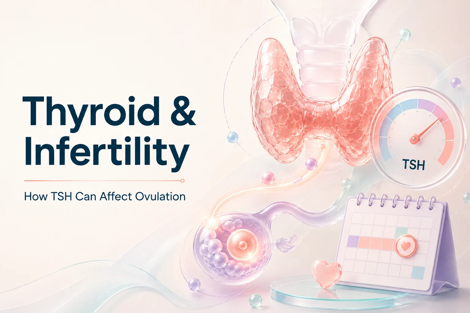
Thyroid and Infertility: How TSH Can Affect Ovulation and Pregnancy Planning
When couples are planning a pregnancy, doctors often evaluate different hormones that play a role in reproductive health. Among these, thyroid hormones are important because they influence metabolism, menstrual cycles, ovulation, and pregnancy preparation. The connection between thyroid and infertility is mainly related to how thyroid hormone imbalance can affect
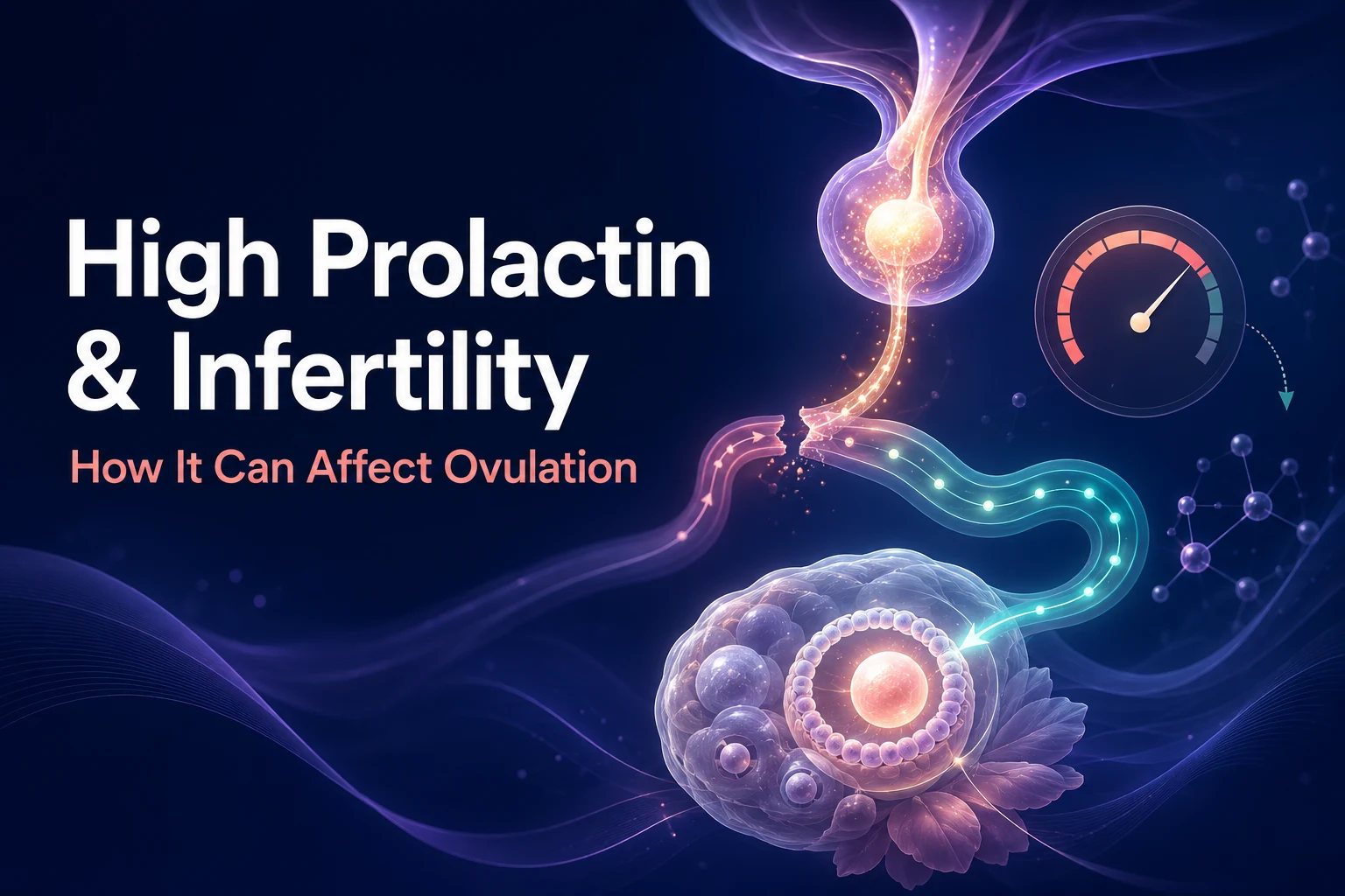
High Prolactin and Infertility: How Prolactin Can Affect Ovulation
When couples are trying to conceive, doctors often evaluate several hormones that influence reproductive health. One of these important hormones is prolactin. While prolactin is best known for its role in breast milk production, abnormal levels can also affect reproductive hormones and ovulation. The connection between high prolactin and infertility